CivArchive
    Medical SEM Style: Corneal Endothelial Cells - v1.0
    Preview 114777053Preview 114777090

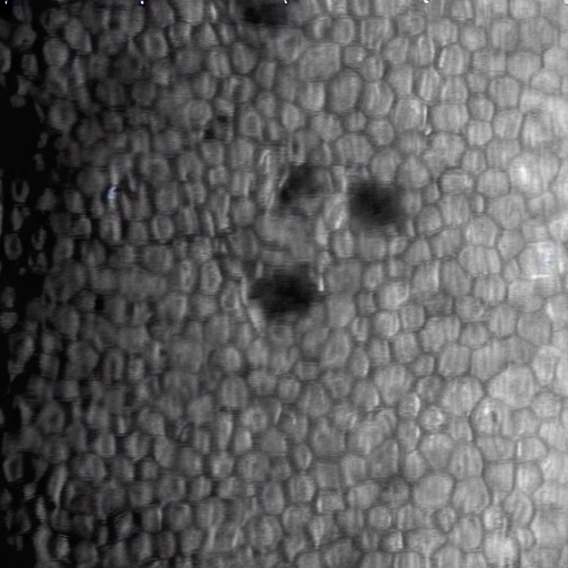
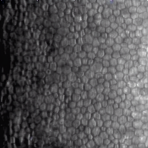
    This LoRA generates photomicrographs of corneal endothelial cells in specular microscopy style (grayscale).

    The model is trained to generate two distinct states:

    1. Normal Cells: Regular hexagonal honeycomb pattern.

    2. Cells with Guttata: Characteristic dark spots/drop-like lesions indicative of endothelial stress or dystrophy.

    Recommended Weight: 1.0 Base Model: SD 1.5

    Training Configuration:

    • Dataset: 1,000 corneal endothelial cell photomicrographs.

    • Resolution: All images resized to 512x512.

    • Training Strategy: 40 repeats per image.

    • Batch Size: 2

    Description

    LORA
    SD 1.5

    Details

    Downloads
    19
    Platform
    CivitAI
    Platform Status
    Available
    Created
    12/22/2025
    Updated
    12/27/2025
    Deleted
    -
    Trigger Words:
    a photomicrograph of corneal endothelial cells, gray scale
    a photomicrograph of corneal endothelial cells with guttata, gray scale

    Files

    sample_500.safetensors

    Mirrors

    CivitAI (1 mirrors)